
Cardiovascular Services
Cardiac Stress Test
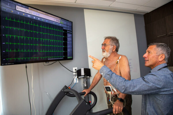
This is an exercise evaluation performed at varying levels of intensity on a treadmill or a bicycle. It monitors your heart rate, blood pressure and cardiac electrical response to warm-up, peak exercise and during rest or recovery.
It is used to detect heart diseases such as blocked arteries in someone who has symptoms such as chest pain or shortness of breath with activity. It is also used to determine someone’s safety prior to engaging in things like a new exercise/training regimen or prior to surgery/anesthesia. It can also be used to determine the effectiveness of treatment for heart problems.
Additionally, exercise testing can monitor one’s fitness level and help design a program to reach a desired fitness level/goal. Imaging such as echocardiography can be added to the stress test (stress echo) to further evaluate cardiac function before and in response to exercise by directly viewing the heart during the test.
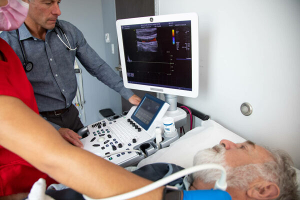
Echocardiography
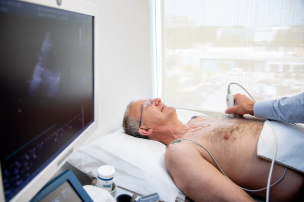
Also known as a cardiac ultrasound or cardiac echo. This test uses sound waves (ultrasound) to create a real-time movie of the heart beating. It’s a painless test that uses an ultrasound probe held against the chest wall to obtain the movies. Ultrasound gel (clear jelly, water-based) is applied to the chest to improve image quality.
Echocardiography is used to detect heart and heart valve function. It can detect areas of damaged muscle, leaky or blocked valves, or small holes or defects that one may have been born with. It is a bedside test that is like an extension of the doctor’s stethoscope.
Vascular Ultrasound
This test uses sound waves or ultrasound to image large to medium sized blood vessels. It can detect blockages or cholesterol build-up in arteries that feed the brain i.e. carotid arteries or abdomen/legs i.e. abdominal aorta.
In addition to detecting blockages, vascular ultrasound can detect enlargement or aneurysms of the aorta that may develop in smokers or those with a family history. Both blockages and aneurysms can be routinely followed by a vascular ultrasound to make sure there is no change or increase in size. If a blockage or aneurysm is significant, early surgical intervention may be warranted to prevent complications such as stroke or arterial tearing (dissection). Carotid ultrasound can also be used to detect early signs of cholesterol buildup that may warrant more aggressive prevention through diet, exercise, and/or medicine.
Electrocardiogram (EKG or ECG)
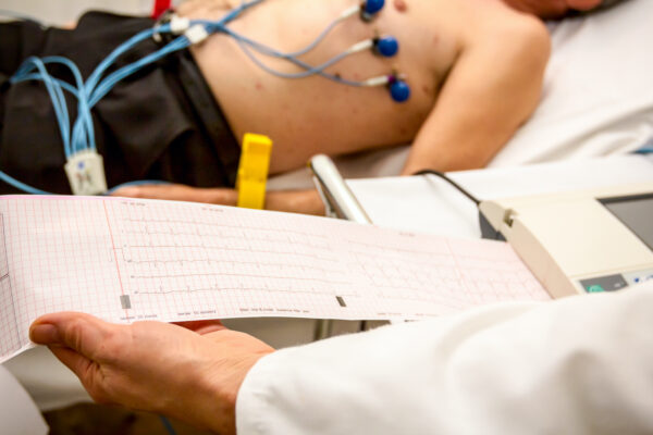
An electrocardiogram (EKG or ECG) is a test that checks for problems with the electrical activity of your heart. An EKG shows the heart’s electrical activity as line tracings on paper. The spikes and dips in the tracings are called waves.
The heart is a muscular pump made up of four chambers. The two upper chambers are called atria. The two lower chambers are called ventricles. A natural electrical system causes the heart muscle to contract. This pumps blood through the heart to the lungs and the rest of the body.
Why is it done? It’s a simple, non-invasive way to check heart’s electrical activity. It can identify the cause of unexplained chest pain or pressure. This could be caused by a blocked coronary artery that can lead to a heart attack. EKG also aids in the evaluation of other symptoms of heart disease such as shortness of breath, dizziness, fainting, and heartbeats that are rapid and irregular. It can detect irregular heart rhythms known as ‘arrhythmia’ such as atrial fibrillation. EKGs can also detect how well medicines are working and see if they are causing side effects that affect the heart or if mechanical devices that are implanted in the heart, such as pacemakers, are working. EKGs should also be performed when other diseases or conditions are present. These include high blood pressure, high cholesterol, cigarette smoking, diabetes, and a family history of early heart disease.
Arrhythmia or Heartbeat Monitors
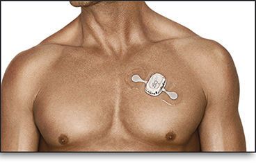
Also known as event recorder, these are battery-powered, wearable devices that either automatically record or you control to record your heart’s electrical activity (EKG) when you have symptoms such as fainting, near fainting, palpitations, rapid heartbeat or extra heart beats.
Current monitors are capable of transmitting tracings over the phone or internet for easy physician review. Some 1-2 day devices require return for readouts. Some devices can be worn for weeks. There are even implantable monitors (about the size of a quarter) that can be used for extended monitoring. Wearing a cardiac event recorder has no risks and causes no pain. However, if you wear electrode patches, the adhesive might irritate your skin. Any skin irritation disappears when the patches are removed.
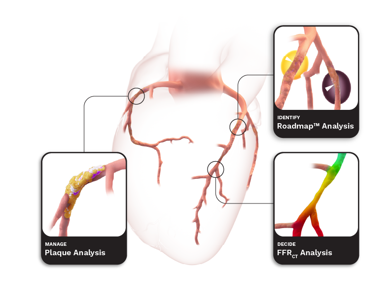
HeartFlow FFRCT
What Is HeartFlow FFRCT?
HeartFlow FFRCT combines CT imaging with advanced AI algorithms to create a personalized, 3D model of a patient’s heart. This model allows our physicians to see how blood is flowing through the coronary arteries and to identify blockages without the need for invasive procedures.
Why This Matters
Heart disease is the leading cause of death in the U.S., and CAD — when arteries that supply blood to the heart become narrowed or blocked — is its most common form. Symptoms like chest pain can signal CAD, but traditional testing methods can be inaccurate or even unnecessary.
Each year, millions of Americans undergo diagnostic tests for chest pain. However, many receive inconclusive results or are subjected to invasive procedures that might not be needed.
With the HeartFlow FFRCT Analysis, we can:
- Improve diagnostic accuracy with the highest-performing non-invasive test available.
- Reduce unnecessary procedures by clearly identifying who does and does not need intervention.
- Enhance patient care with a more personalized and less stressful diagnostic process.
- Clinical trials have shown that using HeartFlow reduces the need for invasive tests by four times, lowers healthcare costs by 26%, and is 75% more likely to pinpoint patients who truly need treatment.